Case: Medial plantar nerve entrapment
Plantar Nerve
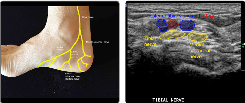
Distal to the medial malleolus, the tibial nerve divides into the medial and lateral plantar nerves.
A slightly more proximal branch, the medial calcaneal nerve, innervates the superficial area of the heel.
Baxters nerve (Inferior calcaneal nerve) is the 1st branch off the lateral plantar nerve. Entrapment of this nerve may simulate plantar fascitis.
Anatomy of the medial ankle, heel and arch nerves.
The tibial nerve bifurcates into the medial calcaneal nerve (posterior) and plantar nerve(anterior).
The plantar nerve then bifurcates into the lateral and medial plantar nerves.
The lateral plantar nerve gives rise to the inferior calcaneal nerve at the posterior arch (this is also called Baxter’s nerve).
Transverse image posterior and slightly superior to the medial malleolus. This is where the medial calcaneal nerve arises posteriorly from the Tibial nerve.
Plantar Nerve
The plantar nerve originates from the tibial nerve at the medial malleolus. The nerve divides into two branches: medial and lateral. It provides motor and sensory innervation to the foot muscles.
- Medial plantar nerve
- Motor fibres: abductor hallucis, flexor digitorum brevis, and flexor hallucis brevis muscles.
- Sensory fibres: sole innervation, 1st, 2nd and 3rd toes, often 4th toe.
- Lateral plantar nerve
- Motor fibres: abductor and flexor digiti minimi, the adductor hallucis, and the interossei muscles.
- Sensory fibres: sole innervation, 5th toe, occasionally 4th toe.

Function
Innervates (sensory and motor):
Terminal Branches:
- Medial Plantar Cutaneous Nerve of Hallux and 3 Medial Common digital nerves
- Proper Digital Nerves
carries sensation from the medial two-thirds of the plantar surface of the foot.
Movements produced:
Flexion and abduction of the big toe (flexor hallucis brevis and abductor hallucis)
Flexion of the toes (flexor digitorum brevis and the first lumbrical muscle)
Pathology/Injury
Medial plantar nerve entrapment:
It is a compression of the nerve branches, where the nerve branches are compressed between bones, ligaments and other connective tissues causing a pain at the inner heel area. Entrapment in the medial longitudinal arch of the foot may result in altered sensation on the medial aspect of the sole of the foot.
Symptoms include almost constant pain whenever adding a pressure to the foot either by walking or sitting, just standing is often difficult.[2] The condition maybe referred to as Jogger's Foot or Medial Plantar Neuropraxia.
Physiotherapy Assessment
Observation:
Local observation for the sole of the foot is the first step of examination, notice any difference compared with the unaffected side, injury or incision, bruises, lump and the skin colour on the related area.
- The atrophy muscle is a sign to indicate if there is an impairment of the nerve that innervates the affected muscle, but it is difficult to be recognised with small muscles.
Palpation:
You can assess the sensation of the areas supplied by the medial plantar nerve and palpate the related area to check any problems relating to the sensation (either hyper sensitivity or impaired sensation) and/or the tenderness degree.
Manual muscle test:
Examine the strength of the muscles that Innervated by the medial plantar nerve, by resisting the movement of the big toe flexion/abduction and toes flexion.
Ultrasound scan plane for the tibial and plantar nerves.

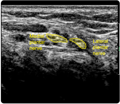
Ultrasound, posterior to the medial malleolus in a transverse plane, sliding the probe distally. The tibial nerve bifurcates into the medial calcaneal (posterior) and plantar nerve(anterior). The plantar nerve then bifurcates into the lateral and medial plantar nerves. At the end of the clip, the probe is rotated slightly demonstrating the lateral plantar nerve in a longitudinal section (direction of Baxter’s nerve).
Tibial nerve bifurcation ultrasound.
Transverse image at the infero-posterior arch of the foot showing the subtle bifurcation of the tibial nerve into the plantar nerves.
The medial plantar nerve is anterior.
The lateral plantar nerve is more posterior and then gives rise to the inferior calcaneal nerve (Baxter’s nerve)
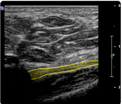
Longitudinal ultrasound of Baxter’s (inferior calcaneal) nerve.
This is immediately inferior to the bifurcation of the tibial nerve into the plantar nerves shown in the previous image.